At Avrupa Sağlık Dental Clinic, we are committed to ensuring our patients have access to the latest dental innovations. Dentistry is evolving at a rate of knots, and we're always on the lookout for incredible technology that can improve the way we work and bring new opportunities to our clients. Intraoral scanning is an advanced form of dental technology, which enables us to carry out 3D tooth scanning and modelling right here in the heart Istanbul.
Intraoral scanners use laser and optical scanning for taking teeth impressions without the long wait and discomfort that conventional scanning methods bring. The scanner makes it possible for dental impressions to be taken without the need to place the gag-inducing putty inside your mouth. This state-of-the-art technology results in an extremely accurate 3D model of your teeth and gums that you will be able to quickly see within the same appointment as the taking of the teeth scans. Avrupa Sağlık Dental Clinic uses this cutting-edge digital impression system to predict the results of dental treatments such as Invisalign, dental implants, dental crowns, and dental bridges.
Benefits of Using An Intraoral Scanner
- No-mess procedure with no gooey mould material placed inside your mouth
- Painless and comfortable, especially beneficial for those who have a strong gag reflex
- Quick impression taking - scanning a full arch of teeth takes only 1 minute to complete
- 3D model of the teeth and gums are created within the same appointment as the impression taking
- Highly accurate impressions obtained, resulting in the creation of extremely precise aligners and teeth restorations
- Provides you with a glimpse of your future smile even before the treatment starts
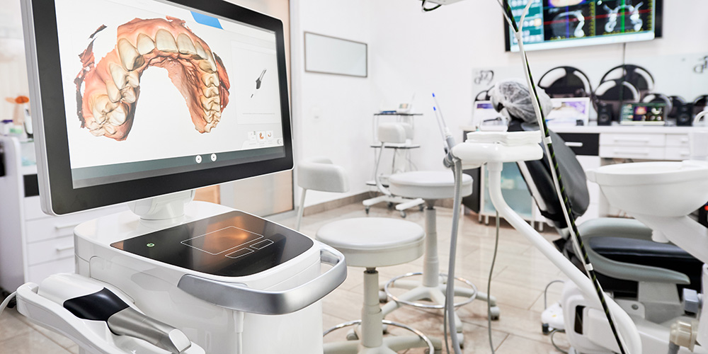
3D Teeth Scanning and Modelling
With intra oral 3D optical scanning, a small wand-shaped device is passed over the teeth to take 3D scans of the teeth. The process is a completely mess free, comfortable and highly accurate method of taking impressions. It is worth explaining that these 'Scans' do not involve any radiation. They are simply thousands of special photographs which are then cleverly stitched together by the software to produce useable 3d images.
The 3D representation on the touch screen monitor allows you to see your teeth from all directions and gain a better appreciation of where the problems are.
We can use the 3D image file to plan how your smile will improve over the course of the treatment by using virtual treatment simulation software. These simulations can be used to illustrate differences possible with various treatment options, giving you the opportunity to help design your treatment outcome.
Dentistry today has advanced leaps and bounds over the last decade. An OPG (Full mouth X-ray) is considered to be a luxury to any private practice due to the high cost factor of the system. In line with our colleagues in the US and UK in which major and minor dental procedure warrant the use of a Panoramic X-ray machine. Its an invaluable tool in proper diagnosis and treatment planning as a lot of pathology that shows on an OPG x-ray is not visible on small X-ray .
A panographic x-ray gives a panoramic view of mouth. It provides valuable information about:
- The position of wisdom teeth, receding bone levels, which is a sign of periodontal disease for Implant surgery
- For jaw-joint problems
- For sinus problems , For Orthodontic Diagnosis
- The Process of X-ray making
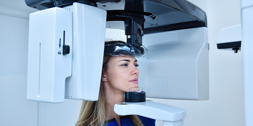
The x-ray film is positioned outside mouth, and the x-ray head rotates around patient. Dental x-rays use high-speed film, so the amount of radiation exposure is very low.
Panographic x-rays are comfortable and safe.
As well been able to take a panoromic x ray of mouth in dental clinics dentists have the facility to take a peri-apical x ray, this enables to focus on a spesific area showing us finer details.
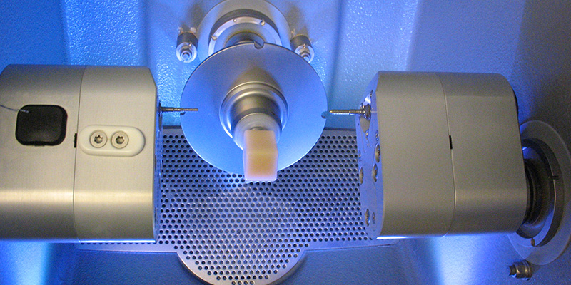
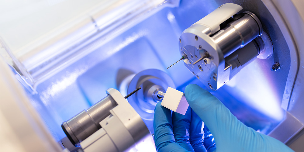
CAD/CAM is revolutionising restorative dentistry, bringing a new level of precision and consistency to the production of crowns and bridges.
Combined with modern materials that offer enhanced biocompatibility and aesthetics, these techniques promote better patient health, giving them the confidence to smile.
The CAD/CAM Process
A six-stage process in which the clinician, technician and CAD/CAM provider collaborate to deliver a high quality restoration.
- Consultation, diagnosis, tooth preparation and impressions
- Model preparation
- Scanning of stone model and framework design
- Framework manufacture (10 year guarantee)
- Porcelain build-up and characterisation
- Fitment of restoration
Difference from conventional restoration
"Chair side" CAD/CAM restoration differs from conventional dentistry in that the prosthesis is typically luted or bonded the same day. Conventional prosthesis, such as crowns, have temporaries placed from one to several weeks while a dental laboratory or in house dental lab produces the restoration.The patient returns later to have the temporaries removed and the laboratory-made crown cemented or bonded in place. An in-house CAD/CAM system enables the dentist to create a finished inlay in as little as an hour in some cases.[CAD/CAM restorations are also more conservative in their preparation of the tooth. As bonding is more effective on tooth enamel than the underlying dentin, care is taken not to remove the enamel layer.
